| Parasite | Kudoa amamiensis |
|---|---|
| Taxonomy | Myxozoa, Myxosporea, Multivalvulida |
| Hosts | Yellowtail (Seriola quinqueradiata), Greater amberjack (Seriola dumerili), Pearl-spot chromis (Chromis notata) |
| Disease name | Kudoosis amami |
| Infection site | Trunk muscle |
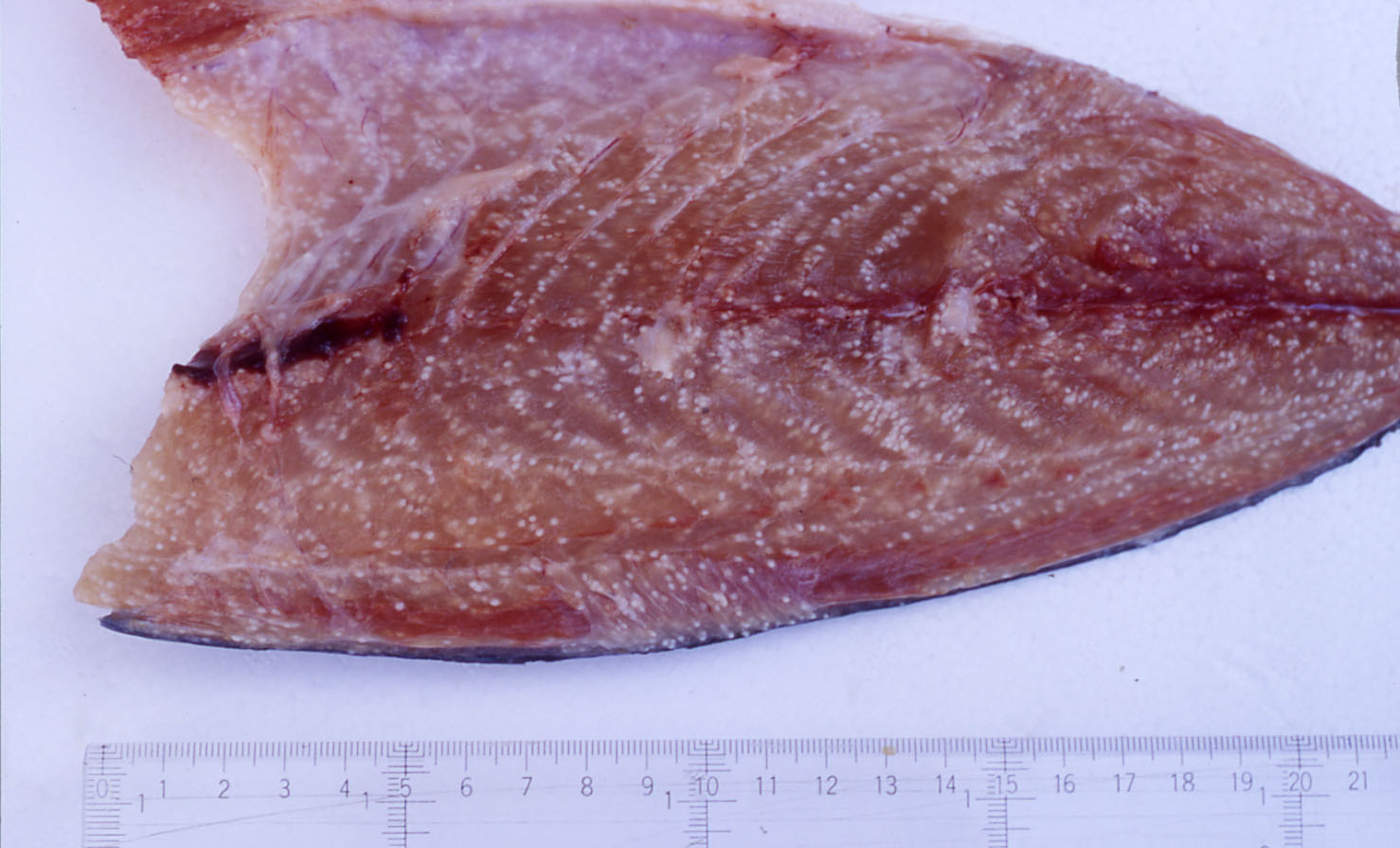
| Clinical signs | Many spherical white cysts (one-several mm) are formed (Fig. 1). It is not fatal to host fish even in heavy infection. |
| Parasitology | A number of spores are produced inside the cyst (Fig. 2). A spore (length 4.5-5 μm;width 5-6 μm) has 4 polar capsules (length 1.5-2 μm).The life cycle is unknown. |
| Pathology | In the early infection period, vegetative stages develop inside the myocytes andremarkable host reactions are not observed. As the plasmodia develop,parasites are encapsulated by fibrous connective tissue, resulting in visible cyst formation. Liquefaction of muscle tissue does not occur. |
| Health hazard | Since this parasite is not infectious to human, it is harmless in food hygiene. |
| Diagnosis | Check the spores by wet-mount of cysts. Kudoa amamiensis can be distinguished from Kudoa iwatai by the spore size and shape (Egusa, 1986). Sample should be smeared and stained by Giemsa or Diff-Quik. The detection methods by IFAT or PCR were developed (Yokoyama et al., 2000). |
| Other information | This disease was noticed in 1975, when yellowtail were reared for exhibition in the Okinawa International Ocean Exhibition (Egusa & Nakajima, 1980). K. amamiensis was first thought to be confined to Amami-Ohshima and a part of Okinawa in Japan, but Whipps et al.(2003) reported the distribution of this parasite in Great Barrier Reef in Australia. The extent of susceptibility to this disease depends on the host fish species, e.g., several hundred of cysts, several dozen of cysts and one or a few cysts (per 1 g of muscle tissue) in yellowtail, greater amberjack and pearl-spot chromis, respectively (Sugiyama et al., 1999). There are no effective methods to prevent this disease. |
| References | Egusa, S. (1986): The order Multivalvulida
Shulman, 1959 (Myxozoa; Myxosporea): a review. Fish Pathol., 21, 261-274. (In Japanese) Egusa, S. and K. Nakajima (1980): Kudoa amamiensis n. sp. (Myxosporea: Multivalvulida) found in cultured yellowtails and wild damselfishes from Amami-Ohshima and Okinawa, Japan. Bull. Jap. Soc. Sci. Fish., 46, 1193-1198. Sugiyama, A., H. Yokoyama and K. Ogawa (1999): Epizootiological investigationon kudoosis amami caused by Kudoa amamiensis (Multivalvulida:Myxozoa) in Okinawa Prefecture, Japan. Fish Pathol., 34, 39-43. (In Japanese) Whipps, C. M., R. D. Adlard, M. S. Bryant, R. J. G. Lester, V. Findlay and M. L. Kent (2003): First report of three Kudoa species from eastern Australis: Kudoa thyrsites from mahi mahi (Coryphaena hippurus), Kudoa amamiensis and Kudoa minithyrsites n. sp. from sweeper (Pempheris ypsilychnus). J. Eukaryot. Microbiol., 50, 215-219. Yokoyama, H., D. Inoue, A. Sugiyama and H. Wakabayashi (2000): Polymerase chain reaction and indirect fluorescent antibody technique for the detection of Kudoa amamiensis (Multivalvulida: Myxozoa) in yellowtail Seriola quinqueradiata. Fish Pathol., 35, 157-162 |
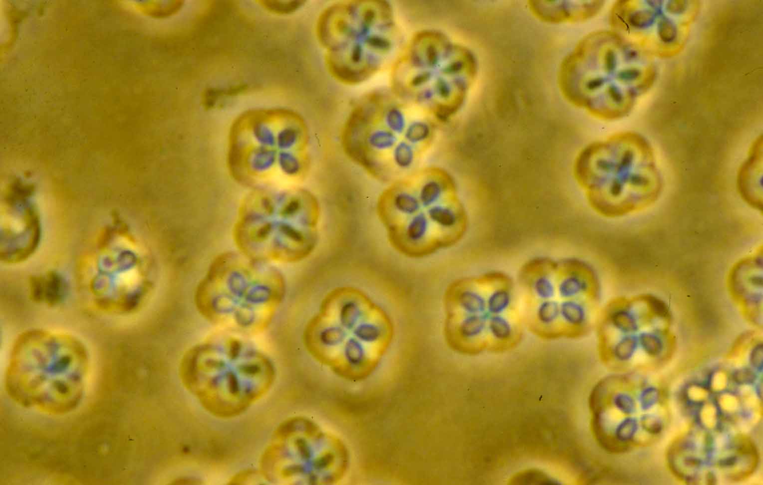
Fig. 2. Fresh spores of Kudoa amamiensis
Fig. 1. A fillet of yellowtail showing Kudoa infection.
